|
Introducing
the Dyop® The
“Revolutionary” Method for Measuring Visual Clarity (Acuity) Helping the world see more clearly, one person at a time.
What Regulates Acuity For vision to be effective and efficient, it needs to be autonomic (so
that we are unaware of that process).
However, acuity is NOT regulated by the brain.
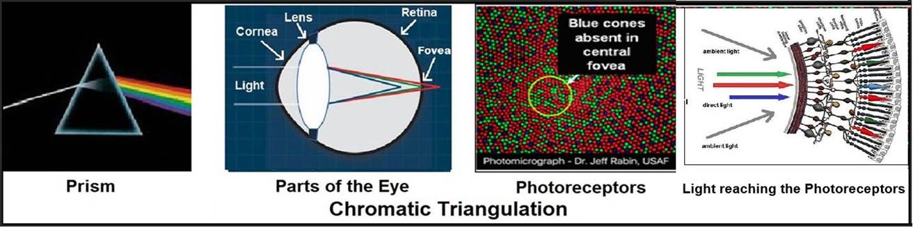
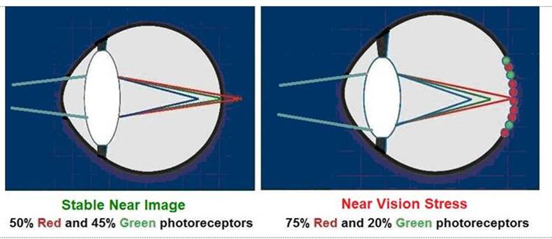
As
light goes through the cornea and lens, it is bent so that Blue is focused in FRONT of the retina, Green is focused ON the retina, and Red is focused BEHIND of the retina. (See the diagram below.) Acuity is regulated by the relative
focal depths and intensity of those colors as they are perceived by the color
sensitive photoreceptors in the fovea at the back of your eyes. Those color sensitive photoreceptors
then send their signals forward to the layer of neuroganglia in
front of the retina. That neuroganglia layer of cells then sends a signal to
the lens to regulate the shape of the lens to bring that image into focus. The process of
combining the response of the color-sensitive photoreceptors to light and
color is like the pixel images you see on
your computer monitor, tablet, or Smartphone. You think you are seeing lines, shapes,
letters, and/or words. What you really are seeing are
pixels of light moving rapidly across the surface of your
computer screen, tablet, or Smartphone in
combinations of Red, Green, and Blue. This process of acuity regulation and
accommodation is called Chromatic Triangulation.
The Chromatic
Triangulation process for acuity/accommodation regulation is based on
the concept of the refraction of light that Isaac Newton discovered in 1665
when he filtered light through a prism. While images are stored in
the brain, acuity is NOT regulated by the brain.
Dyslexia and Color
Perception https://www.dyop.net/documents/Dyslexia_and_Color_Perception-SandraStark.pdf Color Perception as a
Diagnostic https://www.dyop.net/documents/ASOP-06-0651-Dyop_Color_Perception.pdf A simple experiment to demonstrate that
acuity is regulated by the Chromatic Triangulation of Red, Green, and Blue, rather than by the brain, is to close one eye and look around the
room where you are now. You will
notice that with only one eye open you can still determine the relative
distance to nearby objects without the need for binocular vision. = = = = = = = = = = = = = = = = = = = = = = = = = = =
= = = = = = = = = = = = = = = = = = = = = = = = = = How
Acuity is Measured The
properties of visual clarity (acuity) are the SIZE (area) OF THE IMAGE being observed, the VIEWING
DISTANCE to that image, and the
ability of the visual system to PROCESS THAT IMAGE as clearly as possible (Resolution
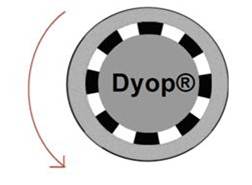
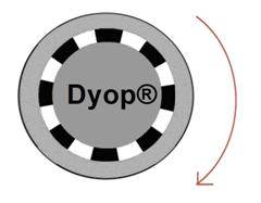
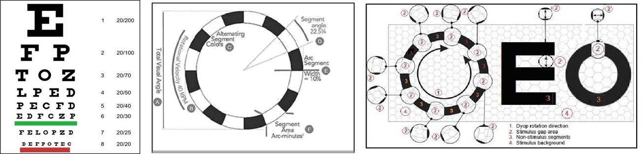
Acuity). As a Dyop® spinning ring gets smaller, the
(equally sized) gaps and segments become so small that it becomes impossible
(sub-acuity) for the eye to detect the spin direction of the Dyop ring rotation. The Dyop acuity endpoint is the smallest Dyop diameter where the direction
of rotation of the spinning ring can still be detected. It serves as a precise, physiological
indicator of visual clarity and vision correction. A Dyop test can measure vision without the need
for patient literacy, measure vision in infants as young as 14
months of age, and let doctors precisely measure vision in
color enabling potential diagnostics for symptoms of dyslexia and glaucoma.
Static acuity tests (such as Snellen
letters) are inherently imprecise, inconsistent.
However, Snellen and other static optotypes mistake the process of Visual Recognition Acuity for Visual Resolution Acuity. The standard Snellen stimulus gap is an overly
large AREA (1.0 arc minutes squared) as the benchmark for vision rather than
the empirically determined smaller Dyop stimulus gap AREA (0.54 arc minutes
squared). Additionally, static vision tests such as Snellen deplete the
dynamic response of the color receptive photoreceptors in the fovea and lack
the uniform precision of Dyop testing.
The result is that static vision tests tend to add excess minus power to acuity and
refractions, lead to angular elongation of the eye
and increased myopia, and indicate that Snellen testing may be a factor in the Global Epidemic
of Myopia. https://www.dyop.net/documents/Snellen_vs_Dyop_Refractions-Sanni.pdf https://www.dyop.net/documents/ASOP-2022-01_Sanni-update.pdf If you wear glasses, a simple test to verify that your lenses are too strong (with too much minus power IF you wear glasses),
is to push your glasses about a half inch away from your face and see if
the words you are reading become larger and more legible. If you notice that the words get more
legible, that Snellen-induced excess minus power of your glasses is typically
about 0.25
to 0.50 diopters. While it isn’t much, it reduces your
cognition and possibly your IQ by 10 points. What becomes even more negative
regarding 21st century Snellen testing
is that the Global Epidemic of Myopia began at almost the same time in human history as the advent of the computer-generated Snellen test. Until then, Snellen letters
were viewed as reflected light with Black
printed letters or shapes on White
paper (to maximize contrast). With the
advent of computers, that contrast pattern was repeated EXCEPT that the printed tests used REFLECTED
light while the computerized tests used EMITTED light. Not only do the static
White Snellen gaps deplete the response and refresh rate of the fovea
photoreceptors, but the overwhelming WHITE background
of the monitor with the Black Snellen letters
further depletes the response of those fovea photoreceptors thus increasing
the addition of refractive minus power, angular elongation and the Global Epidemic of Myopia. https://www.dyop.net/documents/How_Snellen_is_Making_People_Blinder.pdf You can verify that hyper-stimulus visual effect
by briefly staring at a white lightbulb and then closing your eyes. With your eyes closed you should still notice a white stimulus ring for an additional ten
seconds where the photoreceptor response has been depleted. That computerized hyper-stimulus is a contributor to the visual damage
done by using Snellen testing. = = = = = = = = = = = = = = = = = = = = = = = = = = = = = = = = = = =
= = = = = = = = = = = = = = = = = = The Dyop® (Dynamic Optotype™) tests and
concept are covered under U.S. Patent
US 8,083,353 and International
Published Patent WO 2011/022428. for further information contact: Allan Hytowitz at Allan@Dyop.org 5035 Morton Ferry
Circle, Johns Creek, GA, 30022 /
404-281-7798 Copyright ©2025 DyopVision™ Associates. All Rights
Reserved. |